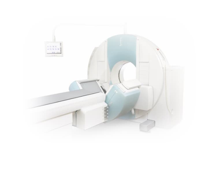
MYOCARDIAL PERFUSION IMAGING


Myocardial perfusion imaging (MPI) is a diagnostic imaging test that is used to assess the blood flow to the heart muscle. It is commonly performed to detect stress and evaluate the heart's function and blood flow. This non-invasive imaging test is crucial in cardiology, helping doctors diagnose and monitor heart conditions.
What Is Myocardial Perfusion Imaging?
Myocardial Perfusion Imaging is a non-invasive procedure that uses a radioactive tracer to evaluate blood flow (perfusion) from the coronary arteries to the heart muscle. The test is done at rest and is usually requested because the ability to exercise on a treadmill is limited.
Small electrode patches are placed on the patient’s chest, which allows continuous ECG monitoring during the test, and intravenous access is established, typically in the veins on your hand. The nuclear tracer (Rubidium-82, Technetium (99mTc) sestamibi) is administered via the vein, and a rest image of the tracer uptake into the heart muscle is obtained using a PET scanner.
The heart will then be “stressed” (exercised) with a pharmacological agent, either adenosine or persantin. The nuclear tracer is re-administered at peak stress to obtain the stress images of the heart muscle. For patients unable to exercise, a pharmacological agent may be used. Single Photon Emission Computed Tomography (SPECT) is also employed to take images of the heart.
The stress images will show areas of the heart muscle with inadequate arterial blood supply due to blockages. This test helps doctors identify areas of reduced blood flow to the heart, which may indicate a blockage or narrowing of the arteries. In Singapore, the MPI ensures patients' heart health is accurately assessed.
How Is a Myocardial Perfusion Imaging Performed?
It is important to prepare properly before undergoing a myocardial perfusion imaging test. You should avoid eating or drinking anything for at least 4-6 hours before the test. 24 hours before the procedure, patients need to avoid any products that contain alcohol, cocoa, such as chocolates and Milo, as well as caffeine, like tea, coffee, and caffeinated energy drinks.
Certain medications may need to be stopped temporarily, so following your doctor's instructions is essential. Stress agents, such as adenosine or persantin, are generally avoided in patients with asthma and respiratory diseases. Generally, there are no side effects of Rubidium-82 and minimal radiation exposure due to its short half-life. A light meal may be recommended a few hours before your appointment. It is also important to inform your doctor if you are pregnant or think you may be pregnant.
During the MPI test, a small dose of a radioactive tracer is injected into a vein, usually in your arm. This tracer travels through your bloodstream and is taken up by the heart muscle. The tracer emits gamma rays, which are detected by a gamma camera that produces heart images. The images show how well blood flows to different areas of the heart. The stress myocardial perfusion aspect might involve an exercise on the treadmill or a medication to simulate exercise conditions in patients who cannot exercise.
Preparing For Myocardial Perfusion Imaging
Preparing for a myocardial perfusion imaging test is essential to ensure accurate results. Your doctor will provide specific instructions tailored to your medical condition, so it is important to follow them closely. Generally, you will be advised to avoid food, caffeine, and certain medications for at least 4-6 hours before the test. A light meal may be required hours after the first part of the test, ensuring comfort and compliance, especially for male patients who may need additional instructions.
In addition, it is crucial to inform your doctor about any medical conditions you have, as well as any medications or supplements you are taking, as some medications may need to be stopped for a short period, so it is essential to follow your cardiologist's instructions regarding their use before the test.
Do's and Don'ts
When getting ready for a myocardial perfusion imaging test, there are a few things you should keep in mind:
- Do follow your doctor's instructions regarding fasting and medication use.
- Do wear loose and comfortable clothing on the day of the test.
- Do get a good sleep the night prior to your test.
- Don't consume any food or drink for at least 4-6 hours before the test.
- Don't consume any caffeine-containing products, such as tea, coffee, or energy drinks, before the test.
- Don't wear any jewellery or metal objects.
What to Expect on the Day of the Myocardial Perfusion Scan?
On the day of your scan, you will be asked to arrive at the testing centre a few hours before the scheduled appointment. You will undergo an electrocardiogram (ECG) to record your heart's electrical activity and monitor your blood pressure and heart rate.
Once you are prepared, the doctor will inject a small dose of a radiotracer into a vein. You may experience a brief sensation of warmth or a metallic taste in your mouth, which is normal.
After the injection, there is a waiting period of about 15-20 minutes to allow the tracer to be absorbed by the heart muscle.
What Happens During the Myocardial Perfusion Scan?
During the test, the gamma camera will capture images of your heart in various positions to assess blood flow to different areas. Your heart rate, blood pressure, and symptoms will be closely monitored throughout the test.
What Happens After a Myocardial Perfusion Scan?
The entire test usually takes about an hour to complete. Once you are done with the scan, you may go about with your daily activities but, ensure to avoid strenuous activities for the day.
Getting a Myocardial Perfusion Imaging in Singapore
Where Can I Get A Myocardial Perfusion Imaging In Singapore
In Singapore, myocardial perfusion imaging tests are typically conducted at specialized imaging centres, hospitals, or cardiology clinics such as The Harley Street Heart & Vascular Centre.
How Much Does A Myocardial Perfusion Imaging In Singapore Cost?
The cost of a myocardial perfusion imaging test in Singapore may vary depending on the healthcare facility and the specific tests performed. Normally, it can cost anywhere from $1500 to $3500. However, checking with your healthcare provider for an accurate cost estimate is recommended.
Who Needs a Myocardial Perfusion Imaging?
A myocardial perfusion imaging is recommended for individuals who may have symptoms of coronary artery disease, such as chest pain, shortness of breath, or fatigue. It is also performed to assess blood flow to the heart in patients who have had a previous heart attack or undergone cardiac procedures like angioplasty or bypass surgery.
Risks
What Are the Risks of Myocardial Perfusion Imaging?
Myocardial perfusion imaging is generally considered safe and carries minimal risks. However, potential risks are associated with the radioactive tracer injected during the test. These risks include allergic reactions and a small risk of radiation exposure. The benefits and risks of the test should be discussed with your healthcare provider. If you are pregnant or think you may be pregnant, please bring this to your doctor's attention, as the test may not be recommended.
Is the Myocardial Perfusion Imaging Test Safe?
A myocardial perfusion scan is considered safe when performed by trained healthcare professionals. The radiation exposure from the radioactive tracer is minimal and within acceptable limits. The benefits of the test in diagnosing and evaluating heart conditions outweigh the potential risks.
Can I Undergo the Test if I Am Feeling Unwell?
If you are feeling unwell on your test day, it is essential to inform your healthcare provider. Depending on your symptoms and medical condition, they will determine whether it is safe to proceed with the test or reschedule it for another time. Your health and well-being are of utmost importance, and it is best to follow the advice of your healthcare provider.
Frequently Asked Questions About Myocardial Perfusion Imaging
Below are some of the most commonly and frequently asked questions about myocardial perfusion imaging (MPI).
What Is a Myocardial Perfusion Scan?
A myocardial perfusion scan, also known as a myocardial perfusion imaging test, is a non-invasive imaging technique that evaluates blood flow to the heart muscle. It uses a radioactive tracer to produce detailed images of the heart, allowing doctors to assess the heart's function and detect any abnormalities or blockages in the coronary arteries.
What Is the Purpose of Myocardial Perfusion Scan?
The purpose of a myocardial perfusion imaging test is to evaluate the blood flow to different areas of the heart and identify any regions with reduced or inadequate blood supply. This information helps doctors diagnose various heart conditions, such as coronary artery disease, and determine the most appropriate treatment options.
Which imaging technique uses a radioactive tracer to assess blood flow to the heart muscle?
The imaging technique that uses a radioactive tracer to assess blood flow to the heart muscle is known as Myocardial Perfusion Imaging (MPI). MPI involves injecting a radioactive substance and then using specialized imaging technology to visualize how blood flows through the heart muscle, helping in diagnosing various heart conditions.
What Can I Expect During Myocardial Perfusion Imaging Test?
During a myocardial perfusion imaging test, you can expect to undergo a series of procedures, including the injection of a radioactive tracer, resting and stress scans, and monitoring of your heart rate and blood pressure. The test may take several hours to complete.
What Imaging Is Best for Myocardial Perfusion?
The diagnosis of coronary artery disease can be made using myocardial perfusion imaging techniques such as SPECT or PET. These tests also provide information on the extent and viability of myocardial tissue.
What are the side effects of a myocardial perfusion scan?
The side effects of a myocardial perfusion scan can include allergic reactions to the tracer, temporary changes in heart rhythm, and mild discomfort at the injection site. Rarely, patients may experience dizziness or nausea. Generally, this procedure is considered safe with minimal risks.
What Are the Potential Risks and Complications of the Myocardial Perfusion Imaging Test?
The potential risks and complications of a myocardial perfusion imaging test are minimal. However, some individuals may experience an allergic reaction to the radioactive tracer or stressor medication or have discomfort or fatigue during the stress portion of the test. It is essential to inform your healthcare provider if you have any concerns or pre-existing medical conditions.
When Will I Know the Results of the Myocardial Perfusion Imaging Test?
The results of a myocardial perfusion imaging test are typically available within a few days. Your healthcare provider will review the images and provide a detailed report with their findings. They will communicate the results to you during a follow-up appointment, discussing any abnormalities detected and recommending further treatment or evaluation if necessary.
Conclusion
Myocardial perfusion imaging or MPI is a comprehensive cardiac test used to detect stress and evaluate blood flow to the heart muscle. This non-invasive imaging technique plays a vital role in diagnosing and monitoring heart conditions. By accurately assessing blood flow and identifying areas of reduced supply, healthcare providers can make informed decisions regarding treatment and management plans. If you believe you may benefit from myocardial perfusion imaging, consult with our cardiologists to determine if this test is right for you.


