VENOUS DIAGNOSTICS
The diagnosis of venous disease is a process of identifying accurately the cause of the high blood pressure in the diseased veins as well as the diseased and healthy veins in the vast network that drains blood back to the heart. At the Harley Street Heart and Vascular Centre, we use a variety of simple and advanced testing techniques, some unique to our vascular diagnostic laboratory, to help make the most accurate assessment and diagnosis for each patient. These include Light Reflex Rheography, FleboLED visualization of veins, Venous plethysmography and Duplex ultrasonography. These are carried out by trained and highly skilled vascular technologists and our vascular specialists at our vascular diagnostic laboratory at Gleneagles hospital.
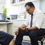
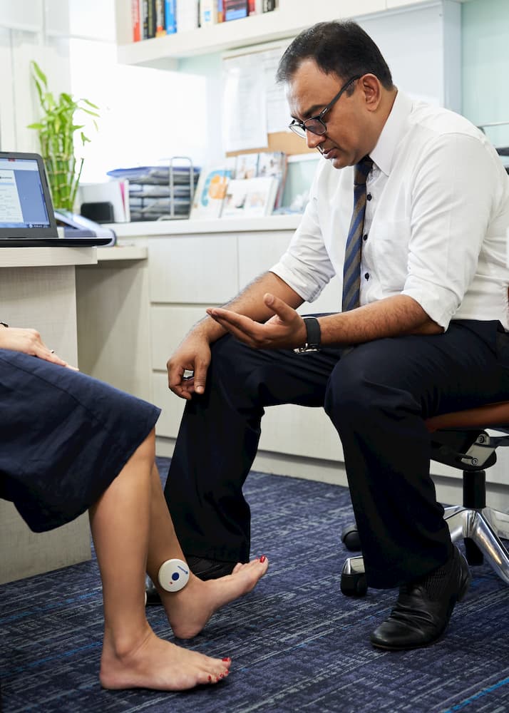
Light Reflex Rheography (LRR)
This rapid non-invasive screening test is used to assess the risk of deep vein thrombosis risk in frequent flyers, in patients with spider veins to rule out any underlying major vein disease and to diagnose whether symptoms of heaviness, aching and swelling in the legs that many people experience are indeed due to any significant vein disease. It is a simple ten-minute test where a wireless pressure probe shaped like a disc is put on each of the patient’s legs and they are then asked to move their move their ankle up and down eight times. Patients who maintain high pressures in the leg despite this ankle movement could have significant vein disease or poor pump function in their calf muscles and may require a further set of vein tests.
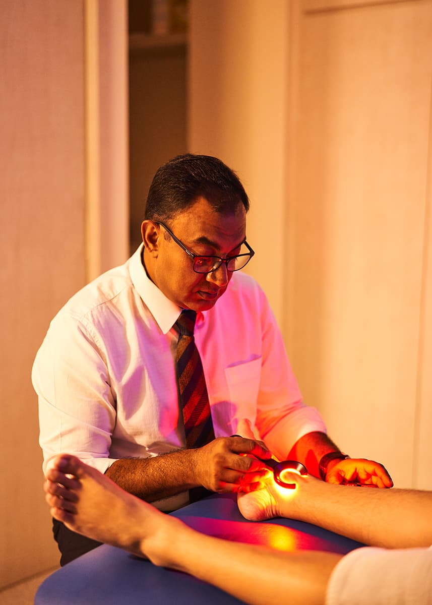
FleboLED Visualization
This test helps to visualize superficial thread veins and spider veins under the skin using 550-660 nm LED lights and map the distribution and anatomy of spider veins. It is used to guide the surgeon towards the most suitable treatment for these veins and identify veins that may be feeding the thread veins but are not visible to the naked eye.
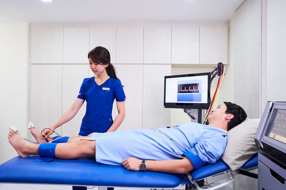
Venous Plethysmography
Plethysmography uses blood pressure cuffs or other sensors attached to an instrument called a plethysmograph (or pulse volume recorder) to measure changes in the volume of a limb (or extremity). Because these volume changes are directly related to the amount of blood flowing through the limb, plethysmography helps to identify problems affecting blood circulation. In the legs, we use plethysmography to identify if there is any obstruction to the return of blood to the heart from the limbs and to quantify how bad the obstruction is.
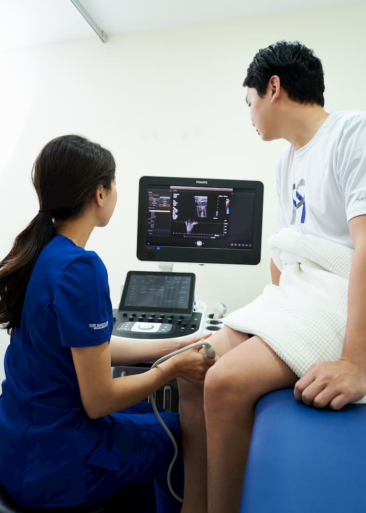
Venous Ultrasound Duplex Scans
These non-invasive ultrasound-based tests are performed to accurately map the extent and severity of disease in the veins. Duplex scans are required for planning the type of procedure that may be most suitable for a patient’s venous condition and to follow-up patients who have had previous procedures.
At the Harley Street Heart and Vascular Centre, our team of vascular specialists and specialist vascular sonographers follow strict and detailed international standard protocols for Duplex scanning and our vascular diagnostic laboratory is a training laboratory for vascular technicians from the United Kingdom and the South East Asian region.