RIGHT HEART CATHETER STUDY
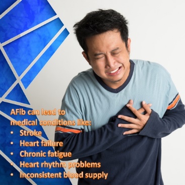
Right Heart Catheter Study is an invasive procedure performed in the catheter laboratory and is used to determine how well the heart is pumping and to measure the pressures in the heart and lungs. This information is valuable in the diagnosis and management of several heart conditions, including heart failure, heart valve disease, congenital heart disease and pulmonary hypertension.
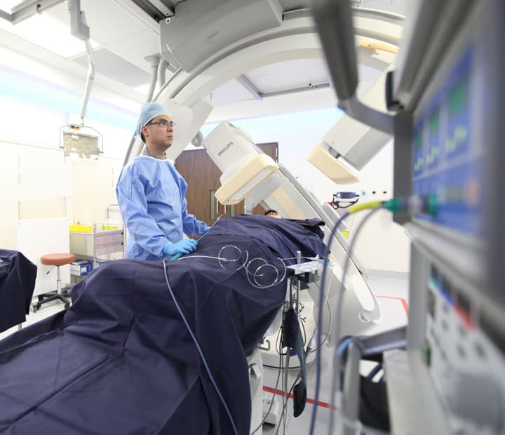
The whole procedure is performed under real time X-ray imaging and requires the assistance of a team comprising nurses and a technologist. An IV line will be started in your hand or arm before the procedure for injection of medication and to administer IV fluids, if needed. You will be connected to an ECG monitor that records the electrical activity of the heart during the procedure using small, adhesive electrodes. Your vital signs (heart rate, blood pressure, breathing rate, and oxygenation level) will be monitored closely. You may be given a mild sedative to help you relax, but no anaesthetic is required.
The cardiologist first places an access tube into the leg vein, then guides a special catheter (a small, hollow tube) to the right side of the heart and passes it into the pulmonary artery, the main artery carrying blood to the lungs. Pressures inside the chambers on the right side of the heart, the right atrium and right ventricle are measured. Indirect measurements of pressures on the left side of the heart are made, as well, by inflating a tiny balloon at the tip of the catheter once the catheter reaches the pulmonary artery. This pressure measurement is called the pulmonary capillary “wedge” pressure (PCWP). The cardiac output, the amount of blood pumped by the heart per minute, is also calculated.
After the procedure, the access tube in the leg is removed and manual pressure applied to close the small wound. You will be expected to lie in bed for a few hours afterwards for routine monitoring, but allowed to eat and drink normally. Usually, if there are no complications, patients are allowed home the same day.