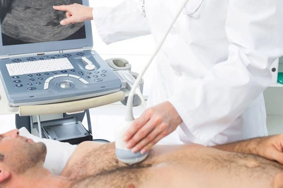
2D Echocardiography

2D Echocardiography is a non-invasive procedure that utilises ultrasound waves to allow your doctor to see your heart's functions and structures. This procedure uses sound waves to evaluate a wide range of conditions or diseases related to the cardiovascular system.
What Is 2D Echocardiography?
Sound waves allow your doctor to look inside your body without needing to pierce your skin. Highly advanced 2D echo machines produce real-time images of your heart by sending sound waves at a frequency too high to be heard, which bounce off your heart chambers and heart muscles, allowing the evaluation of heart function and structure.
How Does 2D Echocardiography Work?
A probe is moved around the heart area outside your body, and the sound waves it sends bounce back to create images of your heart's wall motion and valves in real time. The information is then recorded within a computer, creating a moving image of your heart’s walls and valves.
With this information, your cardiologist can analyse the condition of your heart structures. This technique is crucial in diagnosing conditions such as heart failure, ventricular septal defect (VSD), and regurgitation of blood through the heart valves.
Types of 2D Echocardiography
Depending on your doctor’s recommendation, you may undergo one of two different types of 2D echocardiography.
Transthoracic Echocardiography (TTE)
Transthoracic Echocardiography (TTE) is the most common of the two types of 2D echo procedures. TTE uses electrodes on your chest, which produce sound waves that provide a moving image of your heart. Your doctor can then review the image to understand your current condition better.
Transesophageal Echocardiogram (TEE)
When your doctor cannot get a clear picture of your body using TTE, they may use a Transesophageal Echocardiogram. Less common than TTE, the TEE method uses an endoscope passed down your throat to take a clearer image of your heart from inside your oesophagus, which is especially useful for examining the mitral valve, aorta, and ventricular septal area. The TEE method doesn’t hurt, and you will be on IV for the duration, although some patients feel minor discomfort. Doctors prefer to use the TTE method, but TEE is recommended if required.
Advantages of 2D Echocardiography
2D Echocardiography provides several benefits for the patient and doctor over other methods. Where possible, it is a preferred method and provides high-quality results.
Non-Invasive Procedure
One of the significant benefits of 2D Echocardiography is its non-invasive nature, providing high-quality, real-time images without the need for Doppler radiation or incisions. For many patients, this helps to reduce the pre-procedure nerves. This means no downtime or recovery time is required afterwards, leaving you free to return to your daily life while your doctor assesses the results.
High Accuracy In Diagnosing Heart Conditions
2D Echocardiography can quickly and easily produce highly accurate results. It offers high accuracy in diagnosing heart conditions, such as ventricular dysfunction, cardiomyopathy, and heart valve issues. Whichever method is chosen, your doctor can detect any structural abnormalities within your body and assess your cardiac functions. With this, you can feel confident that your doctor will better understand your current condition after it is finished.
Easy To Perform And Interpret Results
This procedure is relatively simple for your doctor as 2D Echocardiograms are taken primarily with a 2D echo machine. Other than a few standard procedures for the patient, no special preparations are required. Results are available immediately upon completion of the procedure. Allowing your doctor to begin assessing your condition. With this, you aren’t required to wait a lengthy period to understand what’s happening.
Versatility In Assessing Different Aspects Of The Heart
One of the biggest advantages of 2D Echocardiograms is how much information they provide. As the sound waves give a glimpse into your body, your doctor can understand your condition thoroughly. Blood flow and valve functions, heart size and shape, tissue damage, and scarring. Other procedures may require incisions to be made for equipment to assess this information. Something which most patients are pretty grateful for.
What Can 2D Echocardiography Do?
As discussed already, 2D echocardiography can produce a great deal of information for your doctor. Whichever method is chosen, your doctor can gain deep insight into your heart’s condition and functions. With this information, your doctor can take actionable steps towards improving your condition, providing relief or taking further precautions.
Diagnosis Of Heart Disease
When it comes to heart disease, early detection and diagnosis is crucial. 2D Echocardiography allows your doctor to assess the condition of your heart. Any structural defects or blood flow abnormalities will quickly become apparent. From there, your doctor can diagnose your condition and discuss the next steps.
Assessment Of Cardiac Function And Structure
The ejection fraction is the percentage of your total blood pumped with each heartbeat. With your heart functioning properly, your ejection fraction should be above 50%. A 2D echocardiogram lets your doctor understand your heart's current percentage and valve function.
Monitoring Of Heart Conditions
After the procedure, your doctor can begin to track the progress of any heart diseases you may have. Furthermore, they can understand whether treatments or medication are required or need to be adjusted. Interestingly, 2D echocardiograms can also be used to assess changes to your heart after therapy. Providing you with a clearer picture of your current state.
What Heart Problems Can A 2D Echocardiogram Detect?
2D echocardiograms help diagnose some problems or conditions. If your doctor suspects or knows you have any of the following, they may recommend the procedure.
- Arterial blockages in the heart
- Atrial Fibrillation
- Coronary Artery Disease
- Cardiomyopathy
- Damage from a heart attack
- Heart valve problems
- Heart murmurs
- Pericarditis, an infection of the sac around your heart
- Pulmonary hypertension
2D Echocardiography Procedure
Once your doctor has scheduled a 2D echocardiogram for you, there are several steps you will go through during the procedure.
Transthoracic Echocardiography (TTE) Procedure
- Remove any jewellery or objects that may interfere with the procedure at your doctor's request.
- Remove your clothing from the waist up. You will be provided with a gown.
- Lie on a table or bed.
- Electrode patches will be applied to areas across your chest.
- Your doctor will apply gel to your chest.
- They will then move a transducer (ultrasound wand) across your chest to produce the sound waves.
- You must hold still, allowing the doctor to complete the process.
Transesophageal Echocardiogram (TEE) Procedure
- Remove any jewellery or objects that may interfere with the procedure at your doctor's request.
- Remove your clothing from the waist up. You will be provided with a gown.
- Lie on a table or bed on your left side.
- Electrode patches will be applied to areas across your chest.
- Blood pressure cuffs and a pulse oximeter will be attached.
- You will be given a local anaesthetic to numb your throat.
- An IV will be inserted for sedation.
- The transducer will be inserted via an endoscope into your throat
Preparing For The Procedure As A Patient
With a TTE, you don’t need to take any precautions or make preparations. With a TEE, however, you cannot eat or drink six hours prior to the test. Other than drinking water up until two hours prior to the test. Your doctor will explain if there are any other preparations you must make. But generally, a 2D echocardiogram is quite straightforward.
What To Expect During The 2D Echocardiography Procedure?
The 2D echocardiogram is a non-invasive procedure, so you shouldn’t expect any pain or complications. There can be minor discomfort during a TEE as the endoscope is inserted. Depending on your doctor, the transducer process can be uncomfortable during TTE. However, if your doctor cares for their work, you shouldn’t feel discomfort.
Risks and Limitations of 2D Echocardiography
Regarding procedures, most patients are concerned about the potential risks. However, 2D echocardiograms are painless and non-invasive procedures that drastically reduce or remove any possible risks.
Are There Any Risks Associated With 2D Echocardiography?
Firstly, there are no associated risks with TTE. As a non-invasive procedure, you can be confident that there are no common side effects. With TEE, patients occasionally have a sore throat for three to five days after.
You may experience other side effects including:
- An allergic reaction to the medication
- Aspiration pneumonia
- Blood pressure or heart rhythm problems
- Minor bleeding in your oesophagus
Prior to your procedure, the doctor will talk you through all the possible side effects to ensure you are confident about going ahead with it.
Limitations Of The Procedure
If TTE is not viable or doesn’t work, your doctor may recommend TEE.
When 2D Echocardiography May Not Be Suitable?
As discussed above, the TTE method isn’t always suitable. If there are blockages, your doctor may suggest TEE. Whilst slightly more involved than TTE, TTE is still relatively non-invasive.
Why The Harley Street Heart & Vascular Centre Is The Best Choice For A 2D Echocardiography Procedure?
If you believe you require a 2D echocardiogram and are worried about any of the problems stated above, get in touch with The Harley Street Heart & Vascular Centre today. Our experienced doctors are experts in providing 2D echocardiograms in Singapore. Patient care and aftercare are our top priority, focussing on helping you diagnose, treat and recover from your ailments.
Frequently Asked Questions About 2D Echocardiography
What should I avoid before 2D echo?
If you are having a TTE, there are no precautions or preparations necessary. If you are having a TEE, avoid drinking or eating anything but water up to 6 hours prior to the 2D echocardiography procedure.
How long does a 2D echocardiogram take?
Typically, a TTE procedure takes around 30 minutes to an hour. Where else, a TEE procedure can take around 60-90 minutes.
What is the difference between an ECG and an echocardiogram?
An ECG, or electrocardiogram, records the electrical activity of the heart, highlighting rhythm and heart rate anomalies. In contrast, an echocardiogram uses sound waves to create images of the heart, showing its structure and motion, thus helping in diagnosing physical heart conditions like valve problems.
What disease can 2D echo detect?
A 2D echocardiogram can detect various heart diseases, including heart valve issues, coronary artery disease, cardiomyopathy, and congenital heart defects. It also assesses heart function post-heart attack and evaluates the effectiveness of treatments for heart conditions.
What to do before 2D echo test?
Before a 2D echo test, no special preparation is usually required. However, if undergoing a Transesophageal Echocardiogram (TEE), you should fast for six hours prior. Drinking water is permissible up to two hours before the test. It's also advisable to wear comfortable clothing and avoid using lotions or oils on your chest.
Is 2D echo better than ECG?
2D echocardiograms are designed to assess the structure of your heart. ECG’’s look at your heart’s electrical activity.

