CARDIAC COMPUTED TOMOGRAPHY (CT)


Cardiac Computed Tomography or Cardiac Computed Tomography Angiography uses advanced CT technology along with intravenous radio-opaque dye (contrast) to obtain high-resolution 3-dimensional images of the heart. Small electrode patches are placed on the patient’s chest which allows continuous ECG monitoring during the test. The Harley Street Heart & Vascular Centre in Singapore's state-of-the-art facility is equipped with the latest technology and a team of experienced professionals to provide accurate and detailed cardiac computed tomography (CT) scans for the diagnosis and evaluation of various heart conditions.
What is Cardiac Computed Tomography and How Does it Work?
Understanding the Basics of Cardiac Computed Tomography
Cardiac computed tomography, commonly referred to as cardiac CT scans (CT angiography) or CCTA (cardiac computed tomography angiography), is a non-invasive imaging test that utilises X-rays to create detailed cross-sectional images of the heart and its surrounding blood vessels as well as the flow of the blood to the heart. This advanced imaging technique is instrumental in identifying and diagnosing heart diseases.
How Cardiac CT Scans Aid in Diagnosing Heart Diseases
Cardiac CT, commonly known as a CT scan, play a crucial role in diagnosing heart diseases, including heart failure, by providing detailed images of the coronary arteries and detecting blockages, narrowing, or any other abnormalities that may affect the supply of blood flow to the heart. This allows for early detection and timely intervention of the risk of a heart attack.
Benefits of Cardiac CT in Imaging Coronary Arteries
Cardiac CT imaging is particularly beneficial in assessing the health of coronary arteries, detecting the presence of calcified plaque, and evaluating blood flow to the heart, providing invaluable information for accurate diagnosis and treatment planning.
Why Choose Cardiac CT for Heart Disease Diagnosis?
.webp)
Comparing Cardiac CT with Other Imaging Techniques
When compared to other imaging techniques, cardiac CT technology offers a non-invasive and reliable method for visualising the coronary arteries and identifying plaque buildup or blockages, helping cardiologists make informed decisions for patient care. It is one of the many tests for heart diseases that medical professionals will recommend.
Cardiac CT's Role in Detecting Plaque Buildup in Coronary Arteries
Cardiac CT is highly effective in detecting the presence of plaque in the coronary arteries, including calcified plaque, which can indicate the risk of heart disease and potential blockages, prompting proactive management.
How Cardiac CT Helps Assess the Health of Blood Vessels
By accurately imaging blood vessels and blood flow to the heart, cardiac CT aids in assessing the overall health of the cardiovascular system, providing valuable insights into potential issues such as reduced blood flow or vessel narrowing that may slow a patient's heart rate and affect your heart structure and overall heart over time.
Understanding the Procedure: What to Expect During a Cardiac CT Scan
Preparing for a Cardiac CT Scan: Instructions and Precautions
Before a cardiac CT scan, patients will receive specific instructions for preparation, which may include dietary restrictions, medication guidelines, and information on holding your breath during the scan to minimise motion artefacts.
How Long Does a Cardiac CT Scan Take and What Happens During the Process?
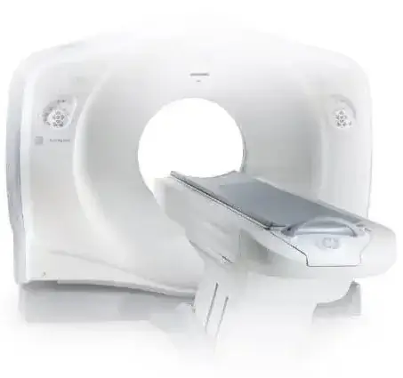
The cardiac CT scan itself typically takes a short duration, during which your healthcare provider will position you on the CT scan. The patient is positioned on the CT scanner while images of the heart and vessels that supply blood are captured. Minimal radiation exposure and the use of contrast dye are part of the procedure.
Possible Risks and Side Effects Associated with Cardiac Computed Tomography
While generally safe, cardiac CT scans may pose minimal risks, including a rare chance of a reaction to the contrast dye used, and radiation exposure. However, the benefits of accurate heart disease diagnosis often outweigh the potential risks.
When Is Cardiac Computed Tomography Recommended?
Indications for Cardiac CT in Various Heart Conditions
Cardiac CT is recommended for various cardiac conditions, including chest pain, suspected coronary artery disease, irregular heart rhythms, and following a cardiac event, to evaluate the condition of the heart and the blood vessels.
Role of Cardiac CT in Screening for Coronary Artery Disease
Cardiac CT serves as an effective screening tool for coronary artery disease, providing detailed imaging of the coronary arteries and detecting the presence of plaque, helping in early intervention and preventive care.
Benefits of Cardiac CT in Evaluating Blood Flow and Heart Function
Assessing blood flow and heart function is an essential aspect of cardiac CT, as it allows healthcare providers to gain insight into the efficiency of blood circulation and the overall performance of the heart, aiding in accurate diagnosis and treatment planning.
Consulting a Specialist: Finding the Right Cardiac CT Imaging Center
Choosing a Reputable Facility for Cardiac CT Scans
When selecting a provider for cardiac computed tomography, it is essential to choose a reputable facility with experienced professionals and state-of-the-art equipment to ensure accurate imaging and reliable interpretation of results for optimal patient care.
Questions to Ask When Selecting a Provider for Cardiac Computed Tomography
Before undergoing a cardiac CT scan, it is important to inquire about the qualifications and experience of the imaging centre's staff, the technology and capabilities of the CT scanner, and the process for interpreting and communicating the scan results.
Importance of Expert Interpretation of Cardiac CT Results
Expert interpretation of cardiac CT results is crucial for accurate diagnosis and effective treatment planning, as experienced specialists can provide detailed analysis and recommendations based on the comprehensive imaging of the heart and blood vessels.
Conclusion
With advances in technology, cardiac CT has become a non-invasive and efficient tool for evaluating cardiac anatomy and function, and it can also guide treatment decisions for patients with heart disease. Additionally, cardiac CT can help identify patients at risk for heart attacks and other cardiovascular events, leading to earlier interventions and improved outcomes.
Overall, cardiac CT has become an important component of modern cardiology, offering a safe and effective means of diagnosing and managing cardiovascular conditions. Its widespread use and continued advancements in technology make it an essential tool for the comprehensive evaluation of cardiac health.
Frequently Asked Questions About Cardiac Computed Tomography
Below are some of the most frequently asked questions about cardiac computed tomography answered by one of our resident cardiologists, Dr. Michael Ross MacDonald.
What Is Cardiac Computed Tomography Angiography (CCTA)?
Cardiac computed tomography angiography, also known as CCTA, is a non-invasive imaging test that uses computerised tomography technology to utilise a piece of special X-ray equipment to produce detailed pictures of the heart, blood vessels that go the heart and arteries that are even close to the heart.
How Does CCTA Differ From Coronary Angiogram?
CCTA is a non-invasive imaging test that uses a CT scanner to obtain images of the heart and blood vessels, while a coronary angiogram is an invasive procedure that involves threading a catheter through the blood vessels into the heart and contrast dye is injected to take X-ray images.
What Is the Purpose of a CCTA?
The purpose of a CCTA is to obtain detailed cardiovascular imaging or images of the heart to diagnose and evaluate coronary artery disease, congenital heart defects, and other cardiovascular conditions.
When Is a CCTA Recommended?
A CCTA may be recommended if a patient has symptoms of heart disease, such as chest pain or shortness of breath. You may need a CCTA to assess the entire cardiac condition of the heart and blood vessels after a heart attack or other cardiac event.
What Is the Role of Calcium in a CCTA?
Calcium scoring is a part of CCTA and it measures the amount of calcified plaque in the coronary arteries, providing information about the risk of heart disease and potential blockages from substances including calcium. Calcified plaque may happen with calcium deposits over time.
Calcium scoring may show a high or low calcium score. However, a high calcium score does not necessarily mean you will experience a heart attack, and a low calcium score does not guarantee immunity from cardiovascular events.
Are There Any Risks Associated With CCTA?
While CCTA is generally safe, there is a small risk of allergic reactions to the contrast dye used, and in some cases, the contrast dye may affect the CT images. Additionally, there is minimal exposure to radiation during the procedure.
Can CCTA Be Performed on Patients With a Pacemaker or Other Implanted Devices?
Patients with pacemakers or other implanted devices may not be able to undergo a CCTA, as the metal in the devices may interfere with the CT examination. However, each case is evaluated individually.
How Long Does a CCTA Procedure Take?
The CCTA procedure generally takes about 10-30 minutes, depending on the type of scanner used and the complexity of the imaging.
What Can I Expect During a CCTA Procedure?
During a CCTA, you will be positioned on the CT scanner table, and electrodes will be attached to your chest to monitor your heart rate. The table will move through the scanner while it takes images of your heart and blood vessels.
Can a CCTA Identify Cardiovascular Disease?
Yes, CCTA can provide detailed images of the heart and blood vessels, allowing for the identification and assessment of cardiovascular disease, including blockages in the coronary arteries and other heart conditions.

