MEDBULLETIN MAY 2025
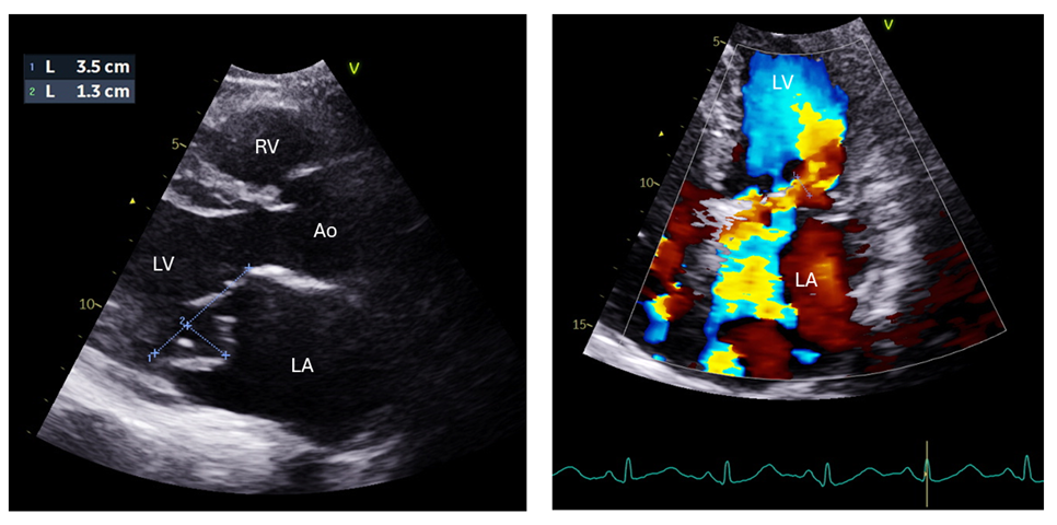
Echocardiogram- parasternal long axis view (left image); four chamber view (right image) with close up of left ventricle (LV) and left atrium (LA). RV= right ventricle, Ao= aorta.
Case Vignette:
A 56-year-old woman with no cardiac history presented to the emergency department with acute shortness of breath. Her symptoms occurred suddenly the night before her presentation and gradually worsened. She was unable to lie flat due to her breathlessness.
On examination, she was tachycardic (heart rate 100 regular) and dyspnoeic (respiratory rate 26) with reduced oxygen saturation on room air (92%). She had bilateral crepitations in her lung fields and a loud pan systolic murmur was heard in her anterior chest wall.
The above are bedside echo images taken at presentation. What do the images show and what is the cause for her acute shortness of breath? What urgent treatment does she need?
Discussion:
Echo findings- the echo images show a prolapsed posterior mitral valve leaflet (P2 cusp) with severe mitral regurgitation (the anteriorly directed yellow/ blue colour jet can be seen in the right-hand image with regurgitant flow from the left ventricle to the left atrium). Her left ventricular function was still normal (LVEF 71%) as the mitral regurgitation was acute onset.
Diagnosis- Acute severe mitral regurgitation secondary to posterior mitral leaflet prolapse (due to a flail leaflet).
Management- the patient was given intravenous Lasix and underwent emergency cardiac surgery the same night of admission. She had a successful mitral valve repair operation and made a good recovery and was discharged one week later..
ANSWERS TO ECG QUIZ
MedBulletin May 2025
MedBulletin Nov 2024
MedBulletin April 2024
MedBulletin Dec 2023
MedBulletin May 2023
MedBulletin Nov 2022
MedBulletin May 2022
MedBulletin Nov 2021
MedBulletin Mar 2021
MedBulletin Sep 2020
MedBulletin April 2020
MedBulletin Sep 2019
MedBulletin March 2019
MedBulletin Sep 2018
MedBulletin March 2018
MedBulletin Sep 2017
MedBulletin March 2017
MedBulletin Sep 2016
2016 Quiz
2015 Quiz