MEDBULLETIN NOVEMBER 2021
Quiz:
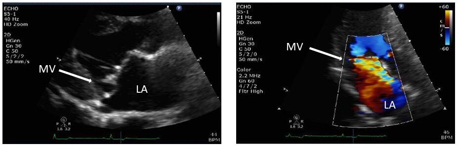
A 50-year-old man who is usually fit and well and cycles 100km every week presented with increasing fatigue and breathlessness over a few weeks. He has no history of cardiovascular disease or any traditional risk factors. Auscultation revealed a pan-systolic murmur in his left sternal region. There were no signs of heart failure and his ECG and baseline blood tests were normal. The echo images are shown in the figure above (parasternal long axis view- MV= mitral valve, LA- left atrium).
Questions:
- What are the main findings on the echo images?
- What additional tests would be useful?
- How would you manage this patient?
Answer:
- What are the main findings on the echo images?
- The echo images show posterior leaflet mitral valve prolapse (left echo image) with an eccentric, anteriorly directed jet of moderate mitral regurgitation (orange/ yellow yet in right image).
- What additional tests would be useful?
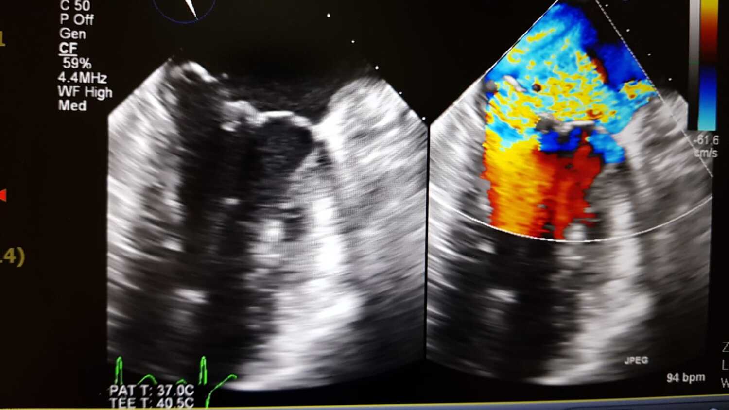
- Transesophageal echocardiogram (TEE) - to assess the mitral valve pathology in more detail and determine the mechanism of the mitral regurgitation. In this case, the TEE showed a flail chord at P2 and P3 with severe mitral regurgitation (worse than shown on the transthoracic echo)- TEE image below.
- A CT coronary angiogram or diagnostic angiogram would be needed prior to any surgical correction of the mitral valve.
- How would you manage this patient?
- Mitral valve surgery – this patient underwent a successful mitral valve repair and made a good recovery. He was back to regular exercise and cycling 3 months’ post-surgery.
ANSWERS TO ECG QUIZ
MedBulletin May 2025
MedBulletin Nov 2024
MedBulletin April 2024
MedBulletin Dec 2023
MedBulletin May 2023
MedBulletin Nov 2022
MedBulletin May 2022
MedBulletin Nov 2021
MedBulletin Mar 2021
MedBulletin Sep 2020
MedBulletin April 2020
MedBulletin Sep 2019
MedBulletin March 2019
MedBulletin Sep 2018
MedBulletin March 2018
MedBulletin Sep 2017
MedBulletin March 2017
MedBulletin Sep 2016
2016 Quiz
2015 Quiz