MEDBULLETIN APRIL 2020
Quiz:
A 56 year old hypertensive male with well controlled diabetes attended his GP surgery with a 3 month history of shortness of breath. Clinical examination revealed mild peripheral edema. He underwent an ECG which showed loss of R-waves across the chest leads. Cardiac MRI was performed pre (a) and post (b) gadolinium contrast.
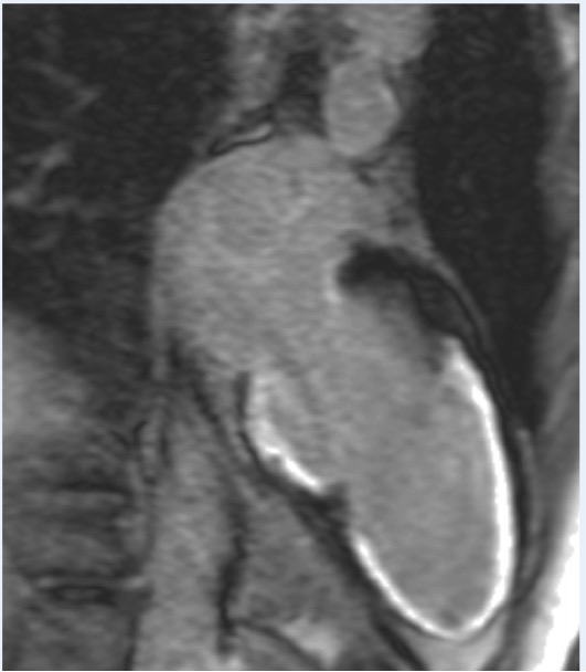
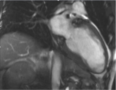
- What do the white areas in image b represent?
- What is the underlying disease process?
Answer:
- The white areas represent previous myocardial infarction. Gadolinium contrast highlights areas of fibrosis. These are subendocardial, and therefore most likely represent a previous myocardial infarction.
- The patient has coronary artery disease. They have had an unrecognised myocardial infarction. Up to 30% of diabetics will have evidence of an unrecognised myocardial infarction on cardiac MRI imaging. This highlights the importance of screening and aggressive risk factor control in our diabetic patients.
ANSWERS TO ECG QUIZ
MedBulletin April 2024
MedBulletin Dec 2023
MedBulletin May 2023
MedBulletin Nov 2022
MedBulletin May 2022
MedBulletin Nov 2021
MedBulletin Mar 2021
MedBulletin Sep 2020
MedBulletin April 2020
MedBulletin Sep 2019
MedBulletin March 2019
MedBulletin Sep 2018
MedBulletin March 2018
MedBulletin Sep 2017
MedBulletin March 2017
MedBulletin Sep 2016
2016 Quiz
2015 Quiz