MEDBULLETIN NOVEMBER 2024
Question:
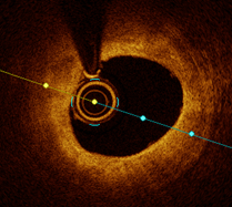
Modern day precision coronary angioplasty involves the use of intracoronary imaging to look at the coronary vessels from the inside. Figure A is how the round imaging catheter (yellow arrow) looks like in relationship to the vessel diameter with its smooth continuous lining of intima (red dots).
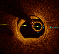
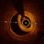
Figure B and C are abnormalities in the same patient that has the normal architecture of the vessel altered. Can you make a guess to what the differences are?
Answer:
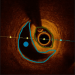
Figure B shows a dissection flap with a break in the continuity in the intima layer of the coronary artery (yellow arrow). This flap extends from the 1 – 9 o’ clock position (yellow line).
Figure C shows the earlier dissection flap entry point leading to a collection of blood (intramural hematoma – black space depicted by the blue dot) in the false lumen (blue outline) and compression the true lumen (yellow outline).
Such dissections can lead to occlusion of the true lumen and compromise coronary artery flow. They generally have to be stented at the dissection entry point to prevent further propagation. These dissections can be spontaneous in nature or during instrumentation in percutaneous coronary interventions and are not always appreciable on just plain angiography. Intracoronary imaging adds another important dimension to current day procedures.
ANSWERS TO ECG QUIZ
MedBulletin May 2025
MedBulletin Nov 2024
MedBulletin April 2024
MedBulletin Dec 2023
MedBulletin May 2023
MedBulletin Nov 2022
MedBulletin May 2022
MedBulletin Nov 2021
MedBulletin Mar 2021
MedBulletin Sep 2020
MedBulletin April 2020
MedBulletin Sep 2019
MedBulletin March 2019
MedBulletin Sep 2018
MedBulletin March 2018
MedBulletin Sep 2017
MedBulletin March 2017
MedBulletin Sep 2016
2016 Quiz
2015 Quiz